X-Ray Imaging
X-radiographic studies provide highly valuable information about elements not visible to the human eye. They reveal details about the artist’s execution technique and the painting’s composition and material history.

Technical Installation
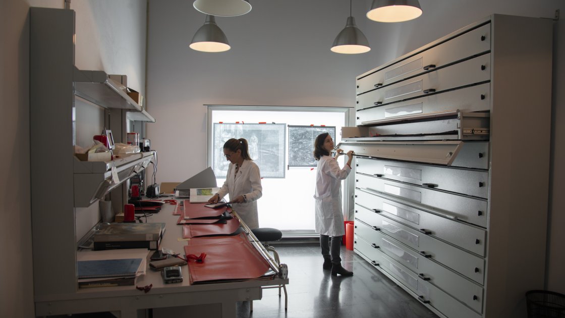
The museum has a restricted area that houses its X-ray equipment. This area consists of two spaces: one for storing and developing X-rays and a shielded enclosure with lead-lined walls to prevent the spread of ionising radiation.
This zone complies with the radiological protection safety protocols established by the Nuclear Safety Council and is staffed by personnel specially trained and authorised to work in facilities of this kind.


We use the following procedure to obtain X-ray images: the artwork is exposed to the X-ray source; the X-ray source emits electromagnetic radiation (X-rays) that is either absorbed by or passes through the object, depending on the material of which it is made and the X-ray parameters used.
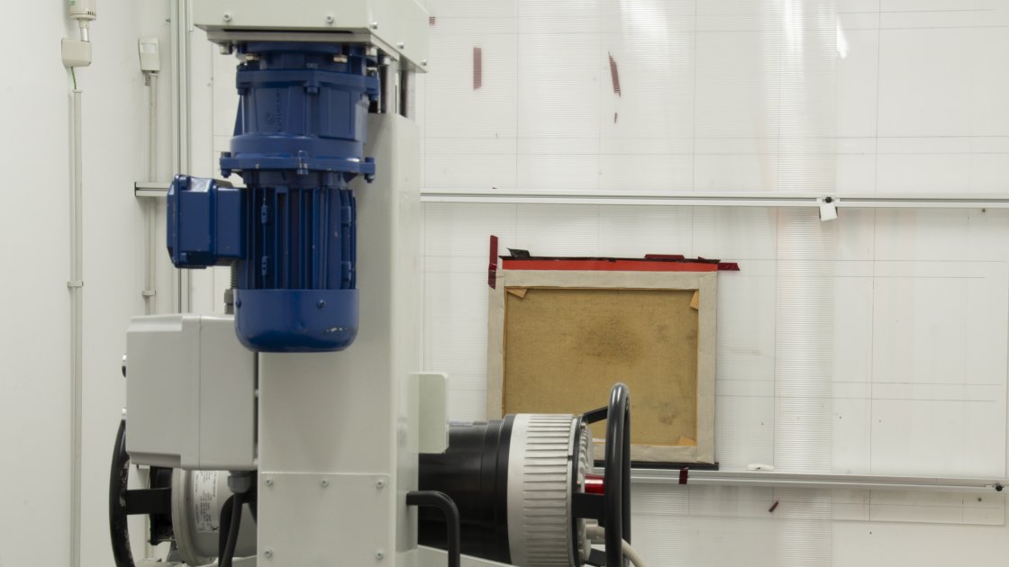
The museum works with an analogue X-ray system. It uses Agfa Structurix D7 X-ray film and industrial X-ray equipment.
A negatoscope is used for the initial viewing of the developed film. For an in-depth analysis, the film is digitised. Digitisation is carried out at the technical documentation department at the Museo Nacional del Prado under a collaboration agreement. The scanner is a Laser Film Digitiser made by Array Corporation. This allows the resulting image to be processed using image editing programmes.
The X-ray film is subsequently developed in a processor, which is located in a dark room, as the film is sensitive to light. For this reason, a red or red-orange safety light is used in these spaces.
As in a conventional photographic laboratory, in order to develop the image, the exposed film has to be immersed in a developing solution that activates the silver halide emulsion.
It is then immersed in a fixing solution and finally washed and dried so that it can be handled, studied and stored. This process therefore requires chemicals, whose residues are stored as waste in suitable containers and collected for processing by specialised companies.
The technical X-ray equipment consists of the following components: X-ray power supply: Izasa 160Kv. X-ray equipment: Yxlon Smart 160 Kv E 0.4. Radiation monitor: Lamse Model RM 1001.


Hidden Results
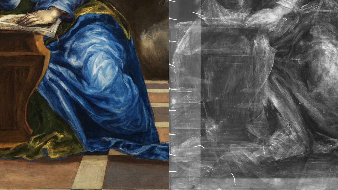
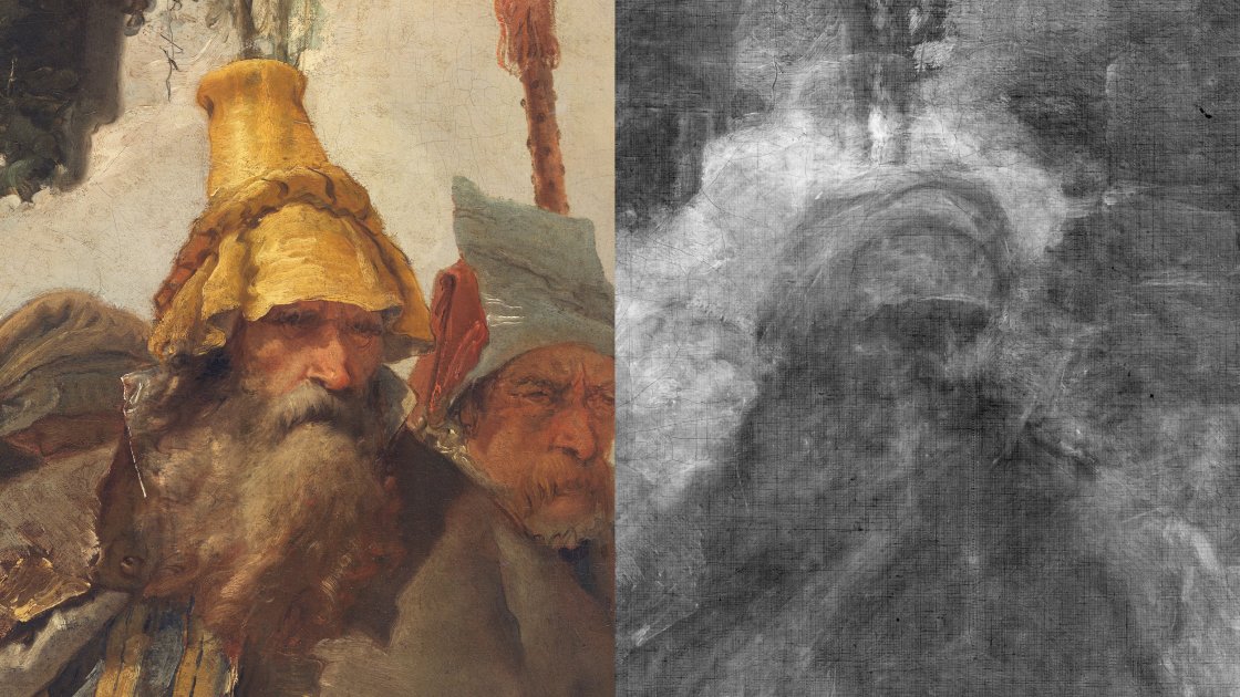
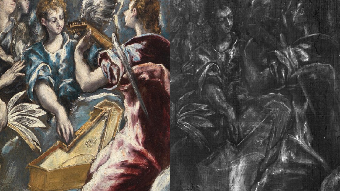
Imaging procedures involving radiation used to analyse artworks provide decisive information about to know the state of conservation and the artist's creative process and technique by studying the elements that define the composition: the form and thickness of the brushstrokes (glazes or impasto) and the method used to apply the paint (brush, palette knife or other) as well as the material characteristics of the components.
The differences in the nature of the material components of the artwork are revealed in an X-ray image by variations in contrast due to the greater or lesser X-ray absorption of the pigments.
X-ray analysis allows us to glean information about the condition of the work: its internal structure, detecting its original and added components, and determining the state of any restoration processes to which it has been subjected, as well as any deterioration.
This technique is decisive for attributing works of art and for detecting compositional changes and pentimenti concealed beneath the layers of paint. It helps discover and show strokes that are part of paintings' initial designs that were subsequently modified or reused.